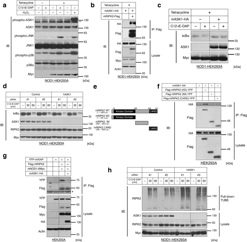
Figure 2.
ASK1 inhibits the activation of the RIPK2 signaling complex. (a) Phosphorylation levels of ASK1 under NOD1 ligand treatment in NOD1-6Myc-stably expressing HEK293A cells (NOD1-HEK293A cells). NOD1-HEK293A cells were stimulated with a NOD1 ligand C12-iE-DAP (1 µg/mL, 30 min). mNOD1 mouse NOD1, H2O2 a positive control of ASK1 phosphorylation, 250 µM, 30 min. *Nonspecific bands. (b) Interaction between RIPK2 and ASK1 in NOD1-HEK293A cells. After the induction of NOD1 with tetracycline, the co-transfected NOD1-HEK293A cells were processed for immunoprecipitation by anti-Flag-tag antibody beads. mASK1 mouse ASK1, mRIPK2 mouse RIPK2. (c,d) Effects of ASK1 overexpression (c) or ASK1 knockdown (d) on NOD-RIPK2 pathway activation. ASK1-overexpressing (c) or ASK1-knockdown (d) NOD1-HEK293A cells were stimulated with C12-iE-DAP (1 µg/mL) for 60 min or the indicated periods of time, respectively. Note that cycloheximide (50 µg/mL) was treated to prevent the rapid feedback synthesis of IκBα. hASK1 human ASK1. (e) Schematic representation of RIPK2 deletion mutants. The numbers indicate the amino acid (a.a.) positions in wild-type (WT). Black rectangles indicate registered domains in the Pfam database (P58801). KD kinase domain, IM intermediate domain, CARD caspase-recruitment domain. (f) Binding ability of RIPK2 domain mutants to ASK1. Co-transfected HEK293A cells were processed for immunoprecipitation by anti-Flag-tag antibody beads. (g) Effects of ASK1 overexpression on the RIPK2-XIAP interaction. Co-transfected HEK293A cells were processed for immunoprecipitation by anti-Flag-tag antibody beads. mXIAP mouse XIAP. *Immunoglobulin G heavy chain. (h) Effects of ASK1 knockdown on K63-polyubiquitination of endogenous RIPK2 under NOD1 ligand stimulation. ASK1-knockdown NOD1-HEK293A cells were stimulated with C12-iE-DAP (1 µg/mL) for the indicated times, from which lysate K63-polyubiquitinated conjugates were purified with a tandem ubiquitin binding entity (TUBE) pull-down assay. Note that superfluous lanes were digitally eliminated from blot images in (c,g,h) as indicated by black lines and the uncropped images are presented in Supplementary Data.

