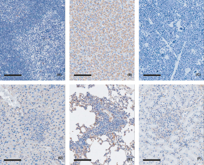
FIGURE 3.

Immunohistochemistry in the lymphoma tissues with a human‐specific anti‐mitochondria antibody in tumorigenicity study. (A) Negative control tissue (spleen), (B) Positive control tissue (U87MG injection site, brain), (C) Lymphoma tissue (spleen), (D) Lymphoma tissue (liver), (E) Lymphoma tissue (lung), (F) Lymphoma tissue (kidney). Human‐specific anti‐mitochondria antibody showed positively stained U87MG tumour cells in brain of the positive control tissue (B). On the other hand, negative results for anti‐mitochondria antibody were observed from the malignant lymphoma tissues (C, D, E, F)
