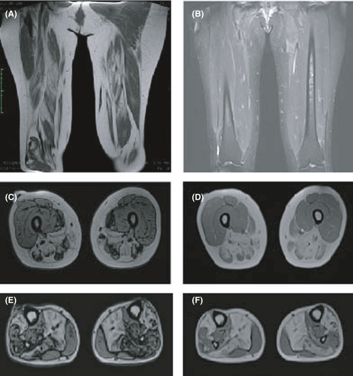
FIGURE 1.

MRI in patient No.5 revealed increased signal intensity of the femur shaft and hamstrings; MRI in patient No.6 revealed pronounced fatty infiltration along with muscle atrophy in the posterior and internal compartments of the thigh muscles and lower leg muscles. (A and B), coronal axial MRI image of thigh muscles in patient No.5. (C and D), transverse axial image of thigh muscles in patient No.6. (E and F), transverse axial image of lower leg muscles in patient No.6
