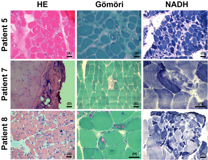
FIGURE 2.

Myopathological changes in patients with GNE myopathy. All specimens were obtained from skeletal muscle. HE staining showed increased fibre size variation (A, G), vacuoles (A), fascial inflammation (D), atrophy (A, G) and regeneration (G). Modified Gömöri trichrome staining showed vacuoles (B) and rimmed vacuoles (E, H) in the fibres. NADH staining showed cores (C, I) and myofibrillar network disarray (F). Scale bar = 50 μm
