FIGURE 3.

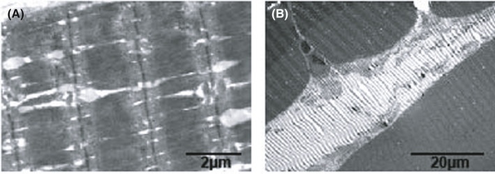
Myopathological changes under electron microscopy in patient No.1 showed atrophic muscle fibres, a dilated sarcoplasmic reticulum and some oedematous and vacuolated mitochondria

Myopathological changes under electron microscopy in patient No.1 showed atrophic muscle fibres, a dilated sarcoplasmic reticulum and some oedematous and vacuolated mitochondria