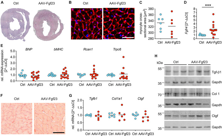
FIGURE 5.
Adeno-associated virus expressing murine Fgf23 mice do not show any signs of pathological LVH. (A) Representative cross-sections of AAV-Fgf23 and Ctrl mice stained with HE (original magnification ×10). (B) Representative cross-sections of AAV-Fgf23 and Ctrl mice stained and wheat germ agglutinin (WGA) (original magnification ×40; scale bar, 50 μm). (C) Quantification of at least 100 individual cardiac myocytes per mouse reveals no size differences between both groups. (D) As analyzed by quantitative real-time PCR, cardiac Fgfr4 mRNA expression is significantly enhanced in AAV-Fgf23 mice compared to Ctrl. (E) The pro-hypertrophic NFAT target genes BNP, bMHC, Rcan1, and Trpc6 are not induced in AAV-Fgf23 mice compared to Ctrl. (F) Representative images of picrosirius red-stained mid-chamber free-wall of AAV-Fgf23 and Ctrl mice (original magnification, ×63; scale bar, 50 μm). (G,H) The mRNA and protein expression of fibrosis-associated markers Tgfb1, Col1a1, and Ctgf and are not altered in AAV-Fgf23 mice. Gapdh serves as the loading control. Data are given as scatter dot plots with means; ***p < 0.001 analyzed using Mann–Whitney test according to D’Agostino and Pearson’s normality test; n = 6–14 mice per group.

