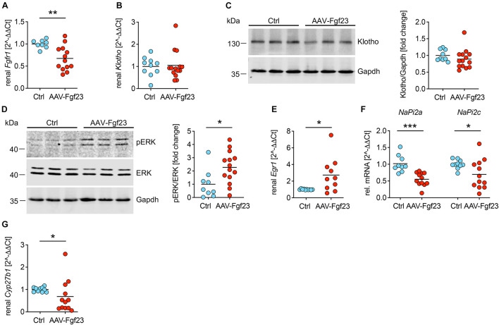
FIGURE 6.
Enhanced secretion of cardiac iFgf23 activates FGFR1/klotho/ERK signaling in the kidney. (A) Analyzed by quantitative real-time PCR, renal expression of Fgfr1 is decreased in AAV-Fgf23 mice compared to Ctrl. (B) Renal Klotho mRNA levels are equal in both groups. (C) Representative immunoblots followed by quantification verify normal klotho protein levels in kidney tissue of AAV-Fgf23 mice compared to Ctrl. Gapdh serves as the loading control. (D) Representative immunoblots in kidney tissue followed by quantification show increased phosphorylation of ERK1/2 in AAV-Fgf23 mice compared to Ctrl with no changes of total ERK1/2 protein. Gapdh serves as the loading control. (E) Renal mRNA expression of Egr1 increases in AAV-Fgf23 mice compared to Ctrl, confirming AAV-Fgf23 mediated activation of ERK signaling pathway. (F) Renal mRNA expression of NaPi2a and NaPi2c decreases in AAV-Fgf23 mice compared to Ctrl. (G) mRNA expression of Cyp27b1 is downregulated in AAV-Fgf23 mice compared to Ctrl. Data are given as scatter dot plots with means; *p < 0.05, **p < 0.01, and ***p < 0.001 analyzed using unpaired t-test or Mann–Whitney test according to D’Agostino and Pearson’s normality test; n = 8–14 mice per group.

