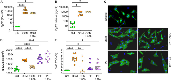
FIGURE 7.
Soluble klotho suppresses cardiac FGF23-induced cardiac hypertrophy in vitro. (A) Stimulation with oncostatin M (OSM) increases endogenous Fgf23 mRNA expression in isolated NRVM irrespective of cotreatment with soluble klotho (sKL). (B) ELISA-based quantification of intact Fgf23 (iFgf23) in conditioned medium of NRVM reveals enhanced iFgf23 release due to OSM treatment, while sKL costimulation does not alter OSM-mediated high iFgf23 secretion. (C,D) Immunofluorescence images of isolated NRVM and quantification of cross-sectional cell area observe hypertrophic growth of OSM stimulated NRVM. Phenylephrine (PE) serves as the positive control for cardiac hypertrophy. Cotreatment with sKL protects from cardiac hypertrophy induced by OSM but not by PE. Myocytes are labeled with anti-α-actinin (green) and nuclei are counterstained with DAPI (blue) (original magnification ×20; scale bar 100 μm). (E) Quantitative real-time PCR analysis shows increased expression of BNP in OSM and PE-treated NRVM compared to Ctrl that is only blocked by sKL in the OSM group. Data are given as scatter dot plots with means; ∗p < 0.05, ∗∗p < 0.01, and ****p < 0.0001 analyzed using one-way ANOVA or Kruskal–Wallis test followed by Sidak’s or Dunn’s multiple comparisons post hoc tests, respectively, according to Shapiro–Wilk normality test; n = 6–9 independent isolations of NRVM; conditioned medium was used from n = 3–7 independent NRVM isolations.

