Figure 1.

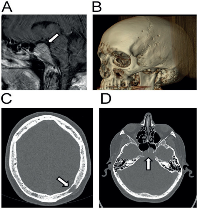
Imaging related to case 2. Sagittal MRI image demonstrating presence of myelomatous mass invading anteriorly from clivus of skull (A). Reconstruction of CT imaging demonstrating multiple lytic lesions of cranial vault characteristic of multiple myeloma (B). Same lytic lesions demonstrated transversely (C). Transverse view of clivus mass invading and compressing the pituitary gland (D).
