Figure 3.

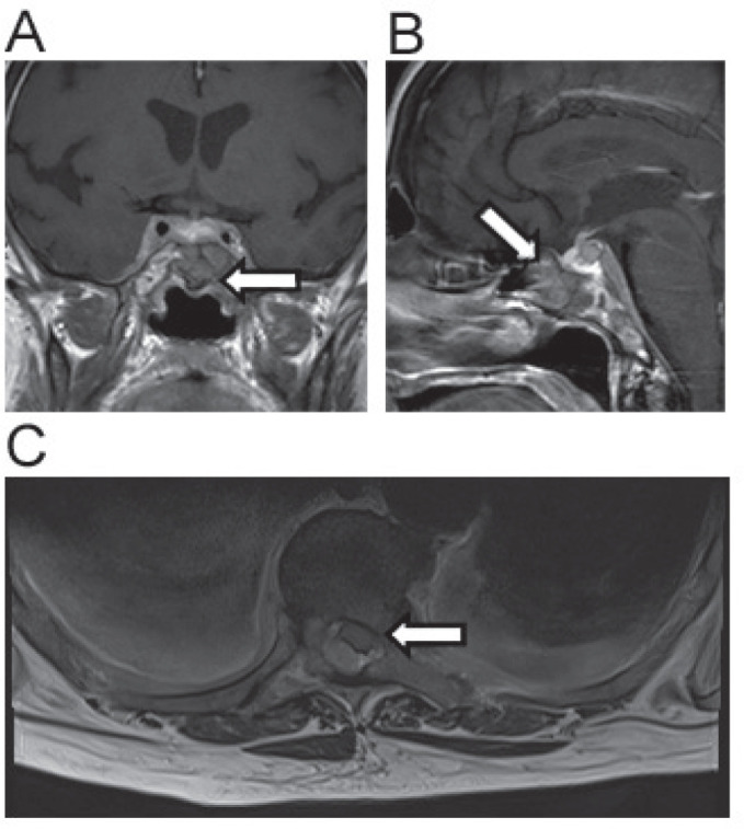
Imaging related to case 4. Coronal MRI image demonstrating compression of pituitary gland by cancer of unknown primary (A). Sagittal view of same lesion (B). Spinal cord compression by a posteriorly situated mass at the level T5/6 (C).

Imaging related to case 4. Coronal MRI image demonstrating compression of pituitary gland by cancer of unknown primary (A). Sagittal view of same lesion (B). Spinal cord compression by a posteriorly situated mass at the level T5/6 (C).