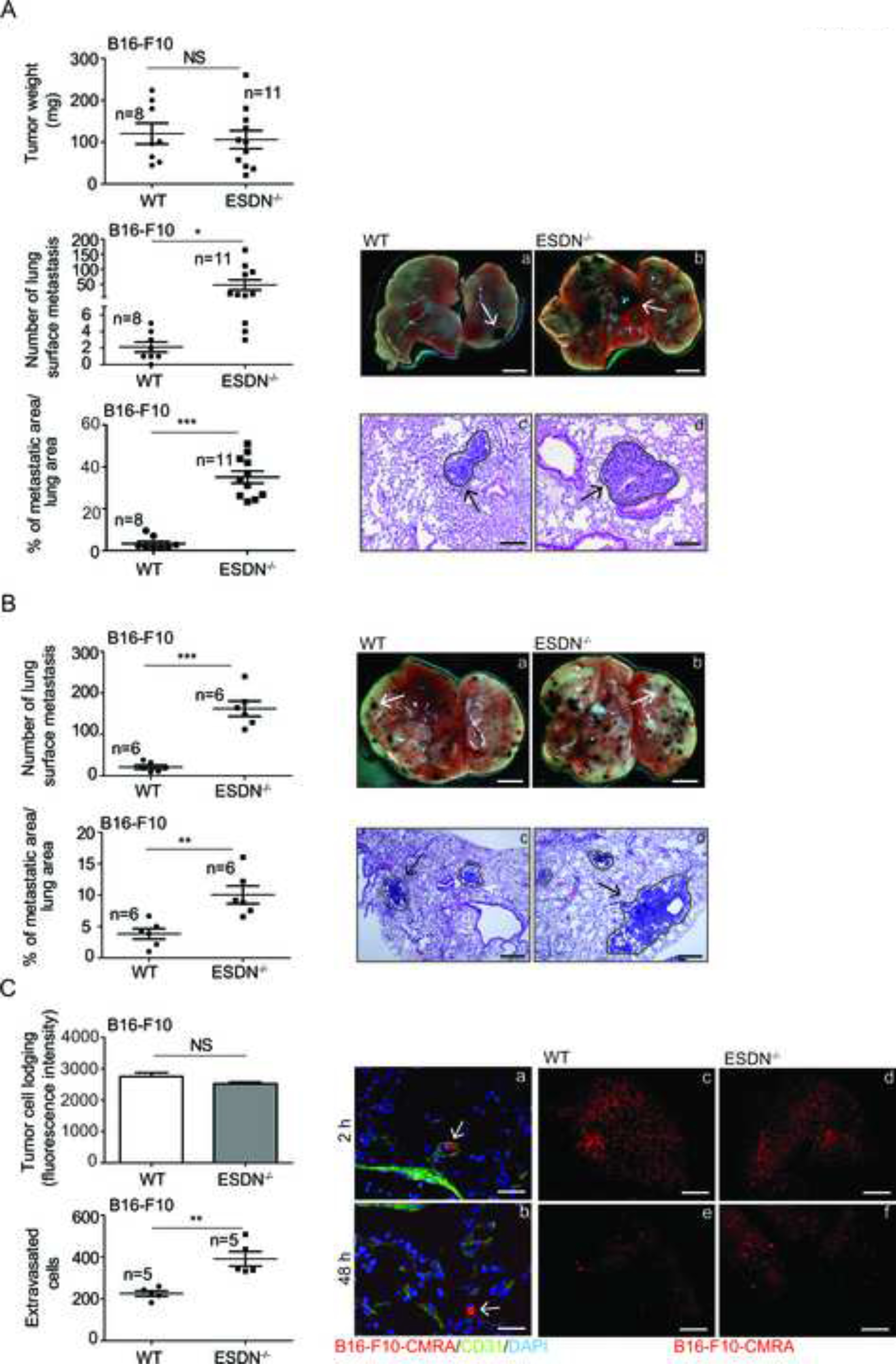
Figure 1 – Stromal ESDN protects from melanoma cell extravasation and metastasis formation but does not affect primary tumor growth in mice.

(A) B16-F10 cells were injected subcutaneously into the flank of syngeneic WT and ESDN−/− mice. Tumors were surgically removed 3 weeks after injections and analyzed; lung metastasis formation was evalutated 3 weeks postsurgery (6 weeks postinjections). Graphs refer to tumor weight (mg), number of lung surface metastasis, or percentage (%) of metastatic areas (24 fields over 6 sections per animal) presented as mean ± SEM. (B) B16-F10 cells were injected into the tail vein of syngeneic WT and ESDN−/− mice. Experimental lung metastasis formation was evaluated 10 days postinjection. (A, B) Representative pictures of the whole lungs (a, b; scale bar = 2 mm) or of H&E-stained lung sections (c, d; bar = 500 μm) are depicted; arrows indicate metastasis formations. (C) CMRA-labeled (red) B16-F10 cells were injected into the tail vein of syngeneic WT and ESDN−/− mice and extravasation was evaluated 2 (a, c, d) or 48 (b, e, f) h later. (a, b) Representative fields of murine lung sections stained for CD31 (green) to highlight blood vessels and counterstained with DAPI (blue). Arrows indicate cells within (a) or outside (b) the vessels; scale bar: 10 μm. (c–f) Representative pictures of whole lungs containing red cells; scale bar: 1 mm. Top graph represents the lodging of injected cells at 2 h as mean of fluorescence intensity ± SEM, whereas bottom graph presents the number of extravasated cells in the whole lungs at 48 h as mean ± SEM. (A–C) Two or three independent experiments were performed and a representative one is illustrated. n = number of animals; SEM = standard error of the mean; H&E = haematoxylin & eosin; WT = wild type; mg = milligrams; CMRA = CellTracker™ Orange; CD31 = cluster of differentiation 31; DAPI = 4’,6-diamidino-2-dhenylindole. * = p < 0.05; ** = p < 0.01; *** = p < 0.001 considered to be statistically significant. NS indicates a nonstatistically significant p-value.
