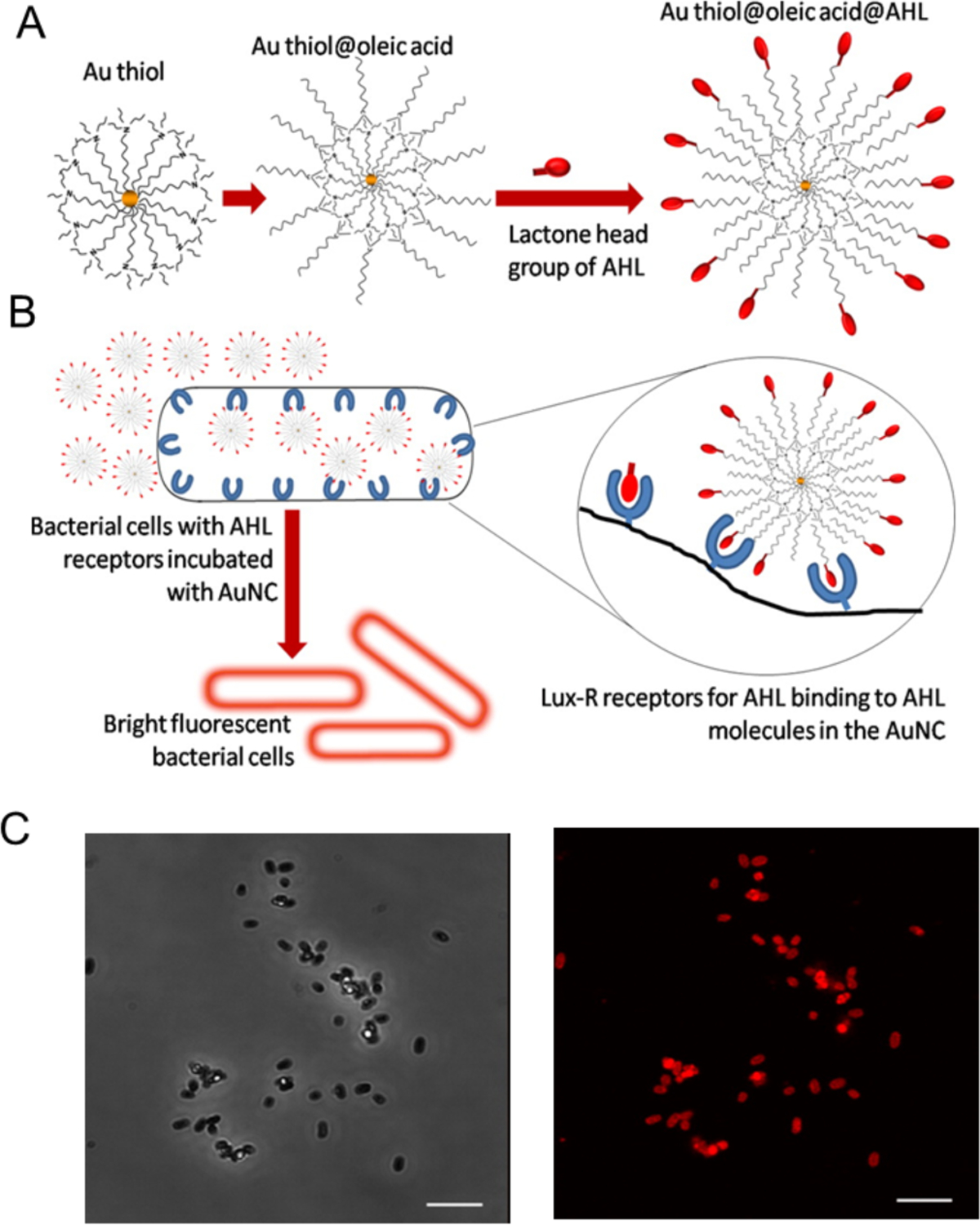
Figure 6.

Fabrication of fluorescent probes for bacterial cells: (A) Structure of the probe with AHL signal molecules deployed on the surface with lactone and amide moieties intact and (B) Specific binding of AHL head groups to receptor sites in Lux-R regulators with bacteria. (C) Confocal microscopy images of E. coli incubated with Au@OA@C8-AHL (AuNC probe functionalized with AHL signal molecules): (top) phase contrast image and (bottom) fluorescence image of the same region. Scale bar is 5 μm. Reproduced from ref.63 with permission from The Royal Society of Chemistry.
