
Figure 1.
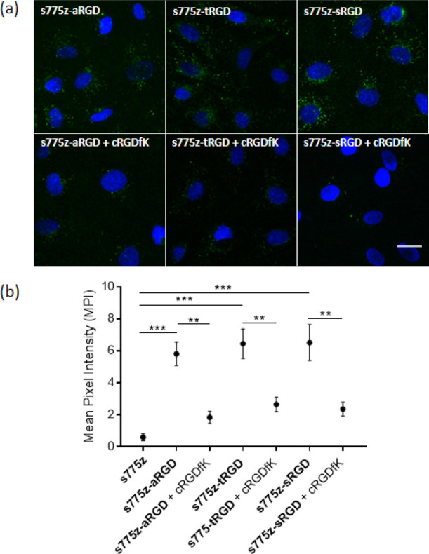
(a) Representative fluorescence micrographs of human A549 lung adenocarcinoma cells treated with 10 μM of one of the targeted probes; s775z-aRGD, s775z-tRGD, or s775z-sRGD. Probe uptake by the cells was blocked by incubating the cells with 200 μM of free cRGDfK for 20 min prior to the addition of the fluorescent probe. In each experiment, probe addition to the cells was followed by cell fixation with paraformaldehyde and treatment with the Hoechst nucleus stain. The probe NIR fluorescence (ex: 769/41 nm, em: 832/37 nm) is pseudo-colored in green for enhanced image contrast and the Hoechst fluorescence (ex: 387/11 nm, em: 447/60 nm) in blue. Length scale = 30 μm. (b) Quantification of cell micrograph fluorescence intensity. ** indicates p < 0.01 and *** indicates p < 0.001.