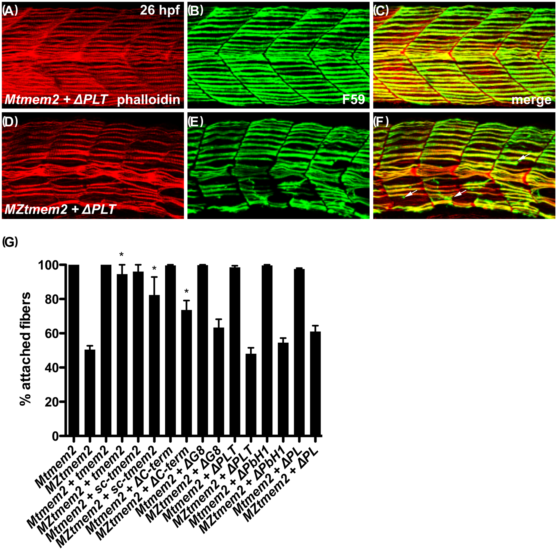
Figure 5. The ectodomain of Tmem2 is critical for promoting skeletal muscle fiber attachment.

Immunofluorescence (A-F) depicts muscle fiber organization, using phalloidin (red) to recognize both fast and slow muscle fibers and F59 (green) to recognize slow fibers; lateral views with dorsal up at 26 hpf. Somite morphology of Mtmem2 sibling embryos is indistinguishable from wild-type, whereas MZtmem2 mutants display muscle fiber detachment (Ryckebüsch et al., 2016). Although expression of full-length tmem2 has been shown to rescue fiber detachment in MZtmem2 mutants (Ryckebüsch et al., 2016), MZtmem2 mutants expressing ΔPLT still display detached fibers (D-F; arrows in F mark examples of detached fibers). In contrast, Mtmem2 siblings expressing ΔPLT exhibit normal fiber attachment (A-C).
Bar graph (G) compares average prevalence of muscle fiber attachment at 26 hpf; error bars indicate s.e.m. In each case, we scored F59+ fibers in 10–11 somites of the left myotome in multiple embryos. Only the F59+ fibers with intact attachments at both boundaries of the somite were scored as “attached”; fibers with dysmorphic attachment, partial detachment, or complete detachment at either end were scored as “detached”, including detached fibers that had retracted substantially. Asterisks indicate significant differences from uninjected MZtmem2 mutants, as determined by Student’s t-test. Full-length tmem2 (p<0.001), sc-tmem2 (p=0.0056), and ΔC-term (p=0.0014) provide significant rescue of muscle fiber attachment in MZtmem2 mutants, whereas other variants do not. Expression of tmem2 variants does not affect fiber attachment in Mtmem2 siblings. Graph includes previously published data for tmem2 and sc-tmem2 (previously called “ectodomain”) (Ryckebüsch et al., 2016). Sample sizes were: Mtmem2, n=4; MZtmem2, n=7; Mtmem2+tmem2, n=6; MZtmem2+tmem2, n=8; Mtmem2+sc-tmem2, n=2; MZtmem2+sc-tmem2, n=5; Mtmem2+ΔC-term, n=3; MZtmem2+ΔC-term, n=5; Mtmem2+ΔG8, n=4; MZtmem2+ΔG8, n=5; Mtmem2+ΔPLT, n=4; MZtmem2+ΔPLT, n=7; Mtmem2+ΔPbH1, n=3; MZtmem2+ΔPbH1, n=4; Mtmem2+ΔPL, n=2; MZtmem2+ΔPL, n=5.
