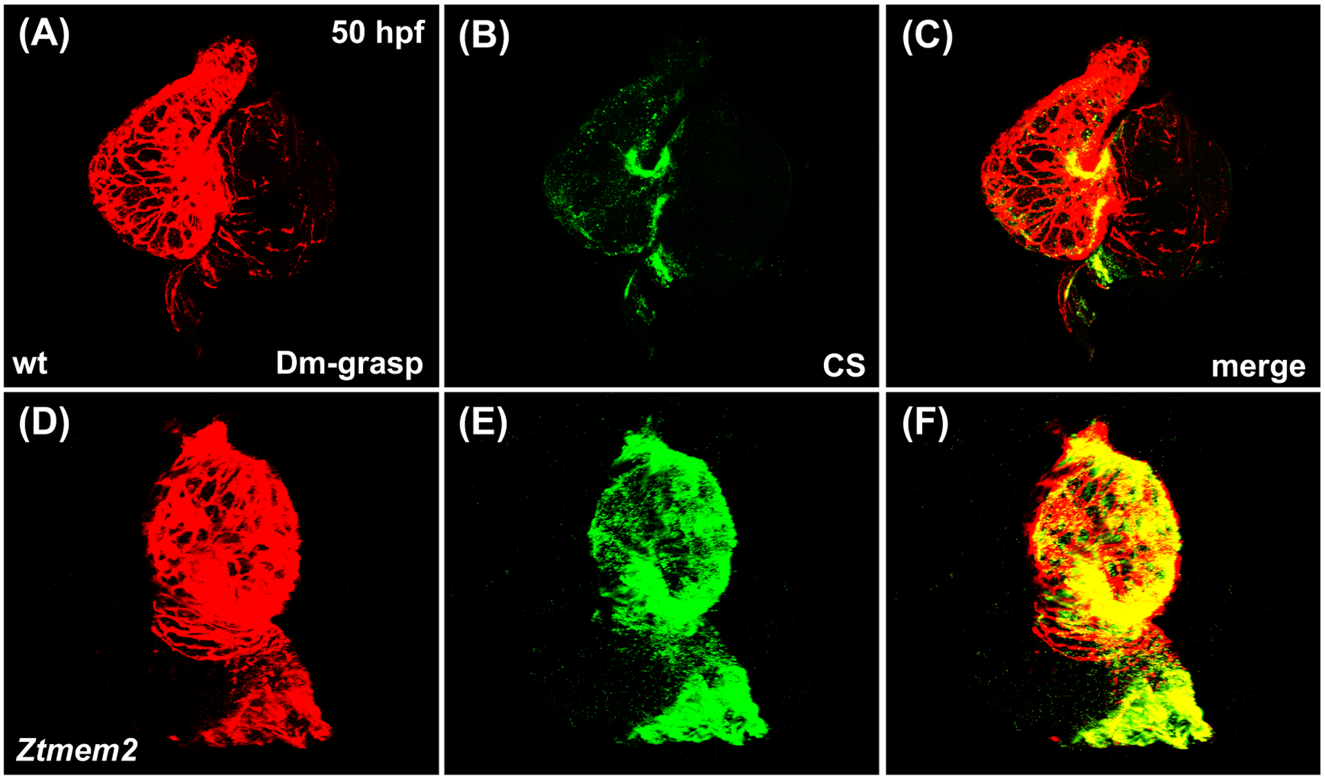
Figure 7. Ztmem2 mutants exhibit excess chondroitin sulfate deposition.

Three-dimensional reconstructions illustrate immunofluorescent localization of Dm-grasp (red) and chondroitin sulfate (CS, green) in wt (A-C) and Ztmem2 mutant (D-F) hearts at 50 hpf. In wt hearts, CS deposition is concentrated primarily within the AVC (B). In contrast, Ztmem2 mutants display increased CS localization throughout the ventricle (E).
