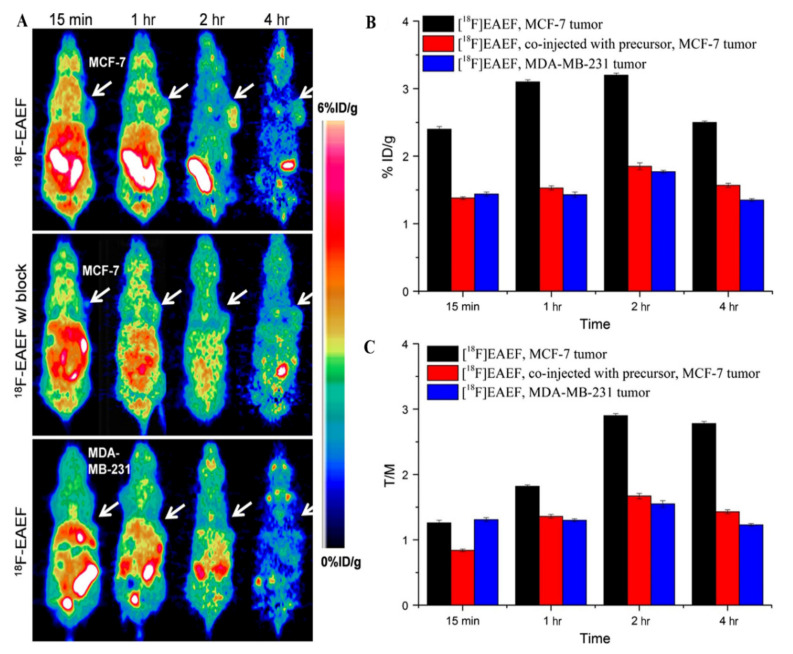
Figure 6.
Chemical structure of PET imaging and tumor uptakes in PR-positive MCF-7 and PR-negative MDA-MB-231 tumor-bearing mice at different times after injection of 18F-EAEF. (A) micro-PET images of MCF-7 tumor with 18F-EAEF (up), co-injected excessive precursor (middle), and MDA-MB-231 tumor with 18F-EAEF (bottom); (B) tumor uptakes and (C) tumor-to-muscle ratios at 15 min, 1 h, 2 h, and 4 h post-injection according to PET imaging [14].

