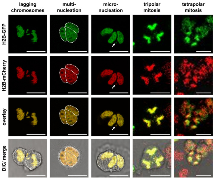
Figure 2.
Changes in the karyotype during HST/PR by lagging chromosomes and multipolar divisions with formation of multinucleation and micronuclei and chromothripsis. Shown are representative images of hybrid cells derived from M13SV1 human breast epithelial cells that were stably transduced with pH2B-GFP (kind gift from Geoff Wahl; Addgene plasmid #11680; http://n2t.net/addgene:11680; RRID: Addgene_11680) or pH2B_mCherry_IRES_puro2 (kind gift from Daniel Gerlich; Addgene plasmid #21045; http://n2t.net/addgene:21045; RRID:Addgene_21045). The plasmids were used to trace green fluorescing GFP-expressing cells and red fluorescing mCherry expressing cells that eventually fuse by forming yellow fluorescing hybrid cells with constitutive expression of both fluorescence genes. Hybrid cells were cultured on chamber slides (ThermoFisher Scientific GmbH, Schwerte, Germany) and images and time-lapse series were recorded using a Leica TCS SP5 confocal laser scanning microscope (Leica, Wetzlar, Germany). Multiple nuclei in multinucleated cells were marked by a dashed line. The arrow points to a micronucleus. Video files of the tri- and tetrapolar cell divisions can be found in the Supplementary Materials Videos S1 and S2, respectively. Bar = 25 µm.

