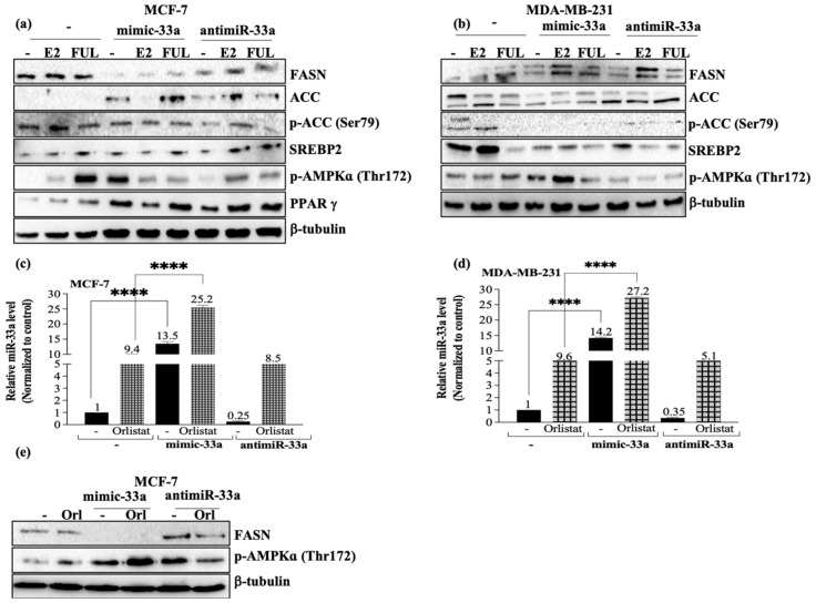
Figure 3.
Investigation of the effect of miR-33a on fatty acid metabolism in MCF-7 and MDA-MB-231 cells. The function of miR-33a expression on fatty acid and energy metabolism in MCF-7 (a) and MDA-MB-231 cells (b) was determined by immunoblotting assay. β-tubulin was used as a loading control. The relative densitometry analysis was performed by two-way ANOVA Tukey’s multiple comparison test (**** p < 0.0001). The miR-33a expression was investigated by evaluating miR-33a in orlistat-treated MCF-7 (c) and MDA-MB-231 (d) cells. Columns represent the mean ± Std.dev of three independent experiments with two repeats (**** p < 0.0001 by two-way ANOVA, Tukey’s multiple comparison test). (e) FASN and p-AMPKα expression in miR-33a overexpressed/suppressed-orlistat-treated MCF-7 cells was investigated by immunoblotting assay. β-tubulin was used as a loading control. The relative densitometry analysis was performed by two-way ANOVA Tukey’s multiple comparison test (**** p < 0.0001). Uncropped Western Blot Figures are shown in Figure S2.

