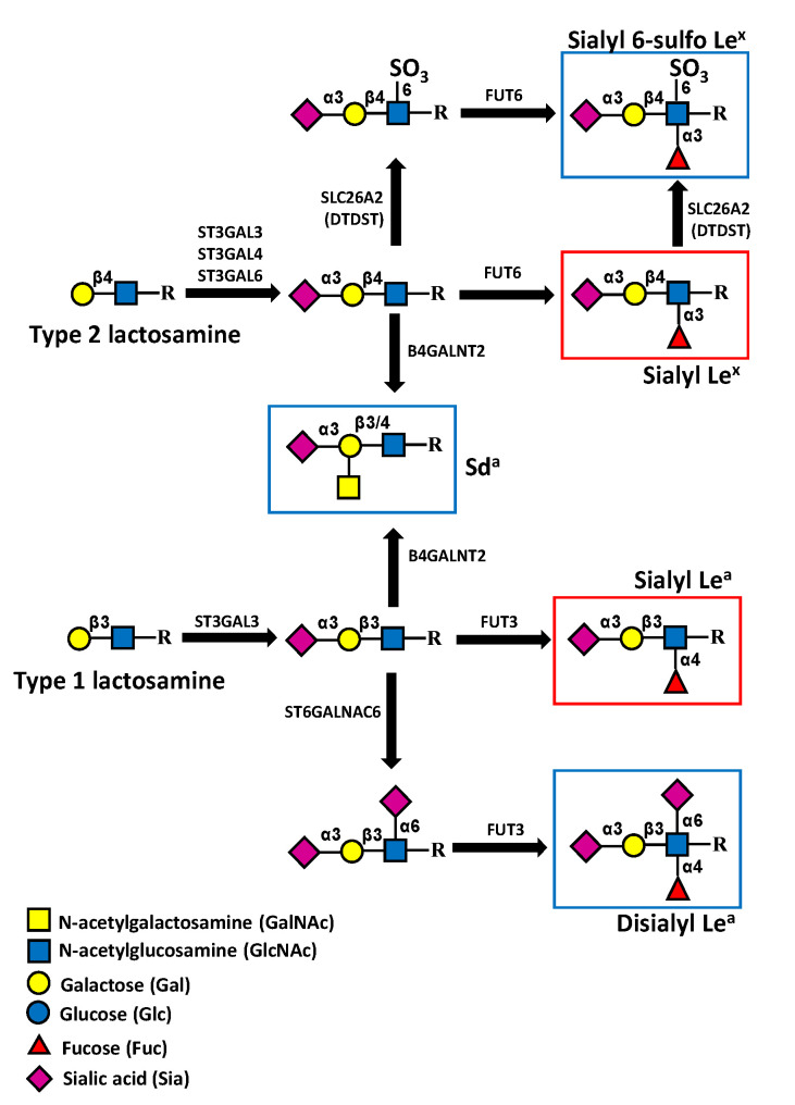
Figure 6.
Sialyl Lewis-related antigens and Sda antigen in colonic tissues. In normal colon, the expression of sialyl 6-sulfo Lex antigen (upper) predominates over that of sLex. However, the reduced expression of enzymes responsible for its biosynthesis, such as SLC26A2 (formerly DTDST) in colon cancer, leads to the overexpression of sLex. The biosynthesis of the di-sialyl Lea antigen (lower), which predominates in normal colon, proceeds from α2,6 sialylation, catalyzed by ST6GALNAC6, followed by α1,4-fucosylation mediated by FUT3. The downregulation of ST6GALNAC6 in colon cancer leads to sLea overexpression. The addition of β1,4-linked GalNAc mediated by B4GALNT2 leads to the expression of the Sda, inhibiting that of sLex and sLea. Cancer-associated structures are boxed in red, while structures associated with normal colon are boxed in blue.

