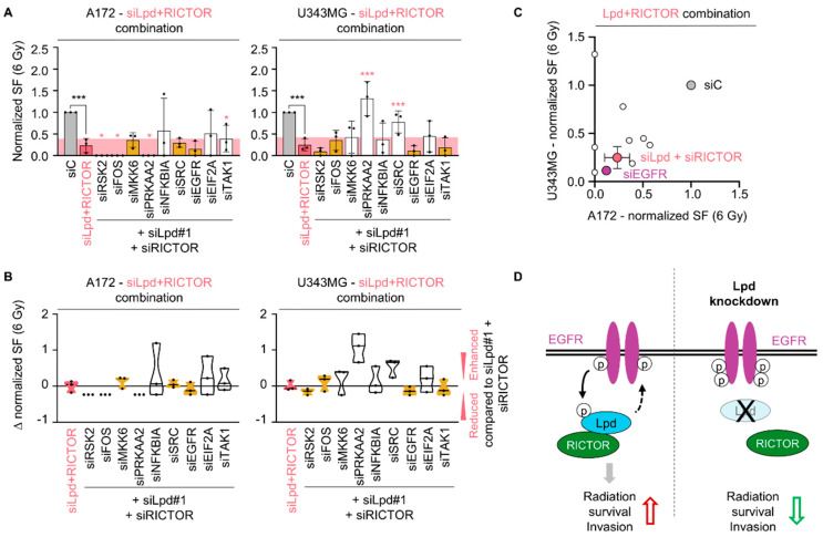
Figure 7.
Lamellipodin and RICTOR jointly promote glioblastoma radiation survival via EGFR. (A) Normalized clonogenic radiation survival at 6 Gy of A172 and U343MG cells upon siRNA-specific triple knockdown of Lamellipodin, RICTOR and indicated proteins. Yellow columns indicate a surviving fraction comparable to double Lpd and RICTOR knockdown, whereas white columns indicate a differing survival. The pink area represents the normalized surviving fraction upon 6 Gy and Lpd and RICTOR silencing. (B) Differences in radiosensitizing effects shown by Δ values of normalized surviving fraction 6 Gy upon knockdown of the indicated proteins subtracted by double Lpd and RICTOR knockdown in A172 and U343MG cells. ∆ value is defined as survival compared to the survival of siLpd and siRICTOR and 6 Gy treated cells: <0 reduced survival, 0 equal survival, >0 increased survival. Yellow color illustrates survival at 6 Gy comparable to Lpd and RICTOR knockdown, whereas white color demonstrates a distinct survival. All results are presented as mean ± SD (n = 3, two-sided Student’s t-test, siLpd + siRICTOR/siC *** p < 0.001, siRNA/siLpd + siRICTOR * p < 0.05, ** p < 0.01, *** p < 0.001). (C) Normalized surviving fraction at 6 Gy of A172 and U343MG shown in (A). (D) Graphical scheme summarizing the interaction of Lamellipodin with RICTOR and the regulation of radiosensitivity and invasion in glioblastoma cells through interplay with the EGFR signaling hub. EGFR exerts a positive effect on Lpd phosphorylation. Conversely, Lpd negatively regulates EGFR levels and phosphorylation. This interplay is cell-line-dependent and may be modulated by basal EGFR activity. Lpd further binds to RICTOR facilitating survival and invasion. Lpd silencing dysregulates EGFR phosphorylation, renders sensitivity to X-rays and reduces invasion. si, small interfering RNA; SF, surviving fraction.

