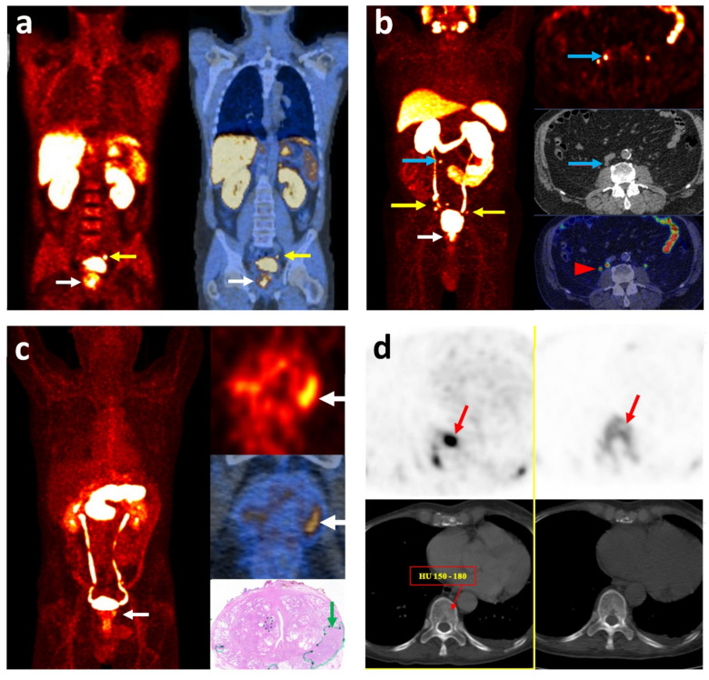
Figure 1.
Imaging with different radiotracers localizing primary prostate cancer and its metastasis. (a) [18F]choline PET/CT: coronal fused images in a high-risk patient with GS = 8 and PSA = 9.6 ng/mL: primary lesion in the prostate gland (white arrow) and a metastatic lymph node in the left iliac chain (yellow arrow). (b) [68Ga]Ga-PSMA PET/CT: MIP (right) and transaxial (left) images in a high-risk patient with GS = 8 and PSA = 4.8 ng/mL: primary lesion in the prostate gland (white arrow) and multiple metastatic lymph nodes in the pelvis (yellow arrow). An unexpected small lymph node is also detected in the upper retroperitoneal region (blue arrow). The red arrowhead shows the physiologic activity in the right ureter. (c) [68Ga]Ga-RM2 PET/CT: MIP (right), transaxial (left) images and pathology section (right lower) in a high-risk patient with pT3a N1 (1/23) and GS = 9: primary lesion in the prostate gland (white arrow). The tumor is outlined with a green line and arrow in the pathology section. (d) Transaxial images of [18F]choline PET/CT (left) and [18F]Na PET/CT (right) in a high-risk patient, showing an early bone marrow metastasis (red arrow) without morphological changes on CT. GS: Gleason Score; MIP: Maximum Intensity Projection; PSA: Prostate-Specific Antigen.

