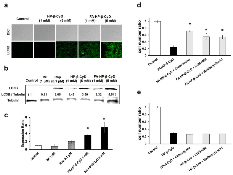
Figure 6.
Induction of autophagy by treatment with FA-M-β-CyD. (a) Detection of autophagy in CML cells. K562 cells treated with FA-HP-β-CyD and HP-β-CyD for 2 h were exposed to Cyto-ID for 30 min. For observation, fluorescence microscopy was used. (b) Effect of HP-β-CyDs on LC3B expression in K562 cells. Cells were treated with medium only (control), FA-HP-β-CyD, HP-β-CyD, imatinib (IM), and rapamycin (Rap) for 2 h. LC3B protein levels were detected by western blotting. (c) The graph shows the fluorescence intensity of the bands. * p < 0.05 compared with the control. (d,e) Effects of chloroquine, bafilomycin A1, and LY294002 on the antitumor activity of FA-HP-β-CyD (d) and HP-β-CyD (e) in BV173 cells. Cells were incubated for 24 h. * p < 0.05 compared with FA-HP-β-CyD.

