Figure 1.
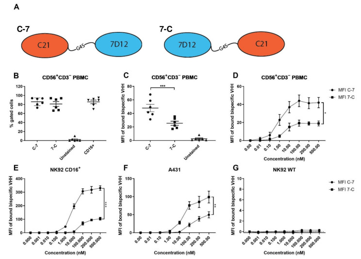
Binding characteristics of C-7 and 7-C bispecific VHHs to CD16 and EGFR. (A) Graphical representation of the bispecific VHHs. (B) Percentage of bispecific VHH+ cells (at 100 nM) and CD16+ cells among CD56+CD3− cells in PBMC, n = 6. (C) Median Fluorescence Intensity (MFI) of bound bispecific VHH on CD56+CD3− cells in PBMC at 100 nM, n = 6. (D) MFI of bound bispecific VHH on CD56+CD3− cells in PBMC n = 3. (E) MFI of bound bispecific VHH to CD16+ NK92 n = 3. (F) MFI of bound bispecific VHH to A431 (EGFR++) n = 8; (G) MFI of bound bispecific VHH to CD16−EGFR− NK92 WT, n = 3. The data are presented as mean ± SEM. Significance is presented as p < 0.05 *, <0.01 **, <0.001 ***. p-values were determined by two-tailed paired t-test (B,C) or two-way ANOVA (D,G). Abbreviations: PBMC = peripheral blood mononuclear cells, C-7 = C21-7D12 bispecific VHH, 7-C = 7D12-C21 bispecific VHH.

