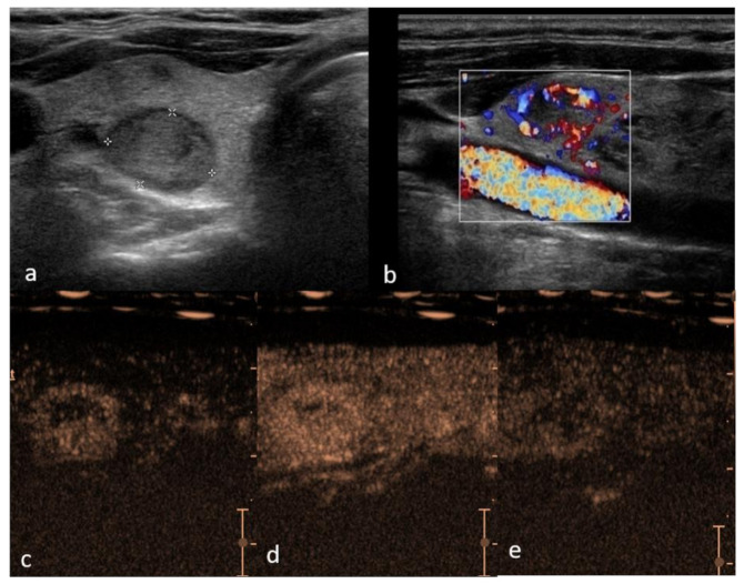
Figure 1.
Right lobe hypoechoic lesion with halo sign, TIRADS 3, Bethesda 2, Follicular hyperplasia (a)—B mode hypo-echogenicity of the structure; (b) color Doppler shows hypervascularity in peripheral part of the lesion; (c) contrast enhancement is predominantly peripheral with smooth ring enhancement, with areas of rapid and intense vascularization in periphery and slow in the center (d) and suggestive slow wash-out (e) in comparison to the adjacent parenchyma.

