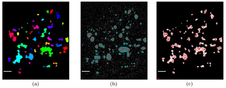
Figure 1.
Example of images of γH2AX clusters after 180 min after 2 Gy irradiation of human skin fibroblasts. (a) Clusters detected by the original program used in [21]. (b) Cluster image obtained with the DBSCAN algorithm (cluster parameter values: Nmin = 110; ε = 200 nm). (c) The same image as (b) but with the closing function applied. The high co-incidence of (a) and (c) can be observed. Scale bar: 2 µm.

