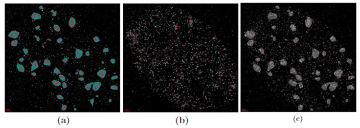
Figure 3.
Examples of cluster images of a human skin fibroblast nucleus 60 min after irradiation with 2 Gy. (a) γH2AX labeling can be found in clusters highlighted by red points in closed areas. (b) MRE11 labeling is dispersed over the cell nucleus and clustered, too. (c) The merged image of (a) and (b) indicating the embedding of MRE11 in γH2AX clusters. Scale bar: 2 µm.

