Figure 9.
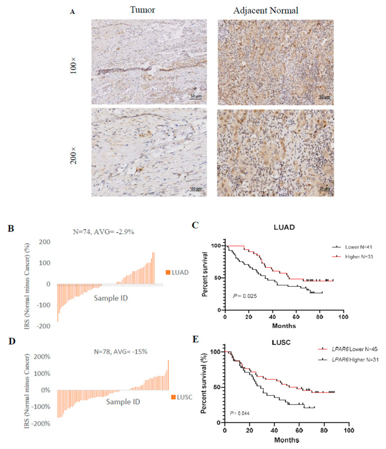
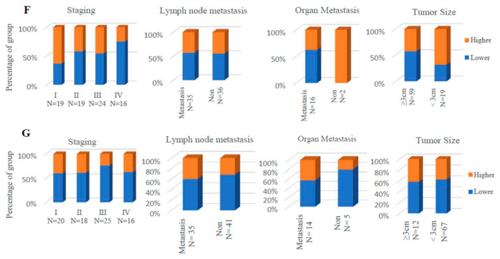
Higher expression of LPAR6 was correlated with clinicopathological parameters in LUAD cohort and was associated with increased overall survival (OS) of LUAD and LUSC patients. (A) Immunohistochemistry staining of the LPAR6 in the tumor and adjacent normal tissues. Red arrows indicated the cytoplasm-stained LPAR6. Bar, 50 μm; (B) The immunoreactive score (IRS) of the cytoplasm LPAR6 staining in 74 paired lung cancer tissues in LUAD cohort; (C) The Kaplan–Meier plot of the OS for lung cancer patients with relatively higher or lower LPAR6 expression levels in LUAD cohort (N = 74; Log-rank test, p = 0.02); (D) The IRS of the cytoplasm LPAR6 staining in 78 paired lung cancer tissues in LUSC cohort; (E) The Kaplan–Meier plot of the overall survival for lung cancer patients with relatively higher or lower LPAR6 expression levels in LUSC cohort (N = 78; log-rank test, p = 0.04). (F,G) The proportion of LPAR6 expression level (higher or lower) in different clinical stages, lymph node metastasis, organ metastasis and tumor size of patients in LUAD (F) and LUSC (G) cohorts.


