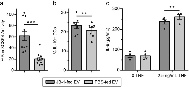
Figure 1.

EV from mice fed with L. rhamnosus JB-1 reproduce in vitro activity of the original bacterium. (a) TLR2 activation by EV was quantified by colorimetric assay using a reporter cell line, with data expressed as a percentage of activity measured for the synthetic TLR2 ligand Pam3CSK4 (300 ng/mL). (b) EV were incubated with BMDCs and subsequent IL-10 expression was measured by flow cytometry. (c) EV were preincubated with T84 cells for 2 h, exposed to 0 or 2.5 ng/mL TNF for 2 h, then IL-8 secretion was measured by ELISA. Error bars represent ± 1 standard error. Each point represents one EV preparation (6–12 mice pooled per preparation). ** p < 0.01, ***p < 0.001
