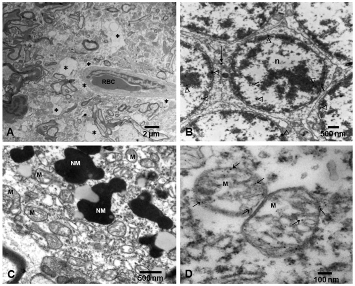
Figure 2.
Electron micrographs of neurovascular unit (NVU) and neural organelles in Metropolitan Mexico City children. (A) Three-year-old boy, substantia nigrae pars compacta showing a capillary with one luminal red blood cell (RBC) surrounded by an extensively vacuolated, fragmented neuropil (*). (B) Cerebellar granular neurons from same child as (A), showing nanoparticles (arrowheads) in intranuclear location and at the membrane interphase between neurons (short arrow). (C) Fifteen-year-old substantia nigrae pars compacta showing numerous mitochondria (M) with abnormal cristae and neuromelanin structures (NM) with nanoparticles. (D) In a closeup, the mitochondria exhibit numerous nanoparticles in the matrix, cristae, and along the double layer mitochondrial wall (arrows).

