Figure 1.
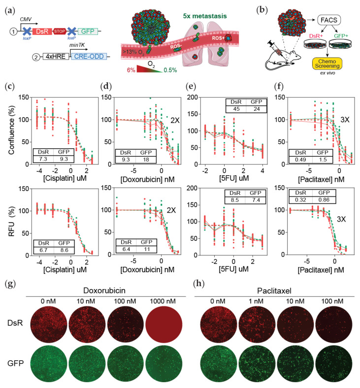
Post-hypoxic cells are less sensitive to doxorubicin and paclitaxel but not 5-FU or cisplatin treatment ex vivo. (a) Lentiviral vectors were delivered to MDA-MB-231 cells to generate a hypoxia fate-mapping system (left). A cartoon depicting that cells that experienced intratumoral hypoxia (GFP+) have a higher probability of forming lung metastases (right). (b) Schematic of the experimental set-up for (c–h). Tumors that formed from the orthotopic injection of MDA-MB-231 hypoxia fate-mapping cells in NSG mice were sorted into DsRed+ or GFP+ populations and then cultured in vitro to determine the IC50 of cisplatin (c), doxorubicin (d), 5-fluorouracil (e), or paclitaxel (f) after 48 h of treatment. IC50 curves were generated by assessing cell viability using the area of the cell culture well covered by nuclear DAPI staining (top) or using a Presto Blue assay (bottom) (N = 3, n = 3); GFP versus DsRed. RFU = relative fluorescence units. (g,h) Fluorescent images of tumor-derived DsRed+ and GFP+ sorted cells treated with doxorubicin (g) or paclitaxel (h) using increasing drug concentrations as indicated. Images are quantified in Figure S1f.

