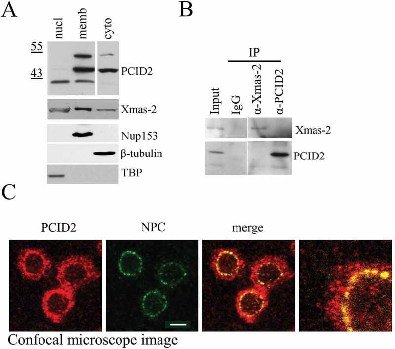
Figure 2.

Characterization of Drosophila PCID2 protein
(a) Western-blot demonstrating the distribution of PCID2 and Xmas-2 in the cell. The same S2 cells were fractionated into nuclear (nucl), cytoplasmic (cyto), and membrane (memb) fractions and 1/10 of each extract was loaded on SDS PAGE. Western-blot was developed with anti-PCID2 or anti-Xmas-2 antibodies. The same extracts were stained with antibodies against Nup153, tubulin, and TBP to confirm the specificity of fractionation (the lower panels). (b) The results of immunoprecipitation experiments with antibodies against PCID2 and Xmas-2 or with IgG (control) from the cytoplasmic extract of S2 sells are shown on Fig. 2a. Blots were stained with antibodies against Xmas-2 and PCID2. (c) Confocal microscope image of the Drosophila S2 cells immunostained with antibodies against PCID2 and NPC and the merged and scaled up images. Scale bar, 10 μm.
