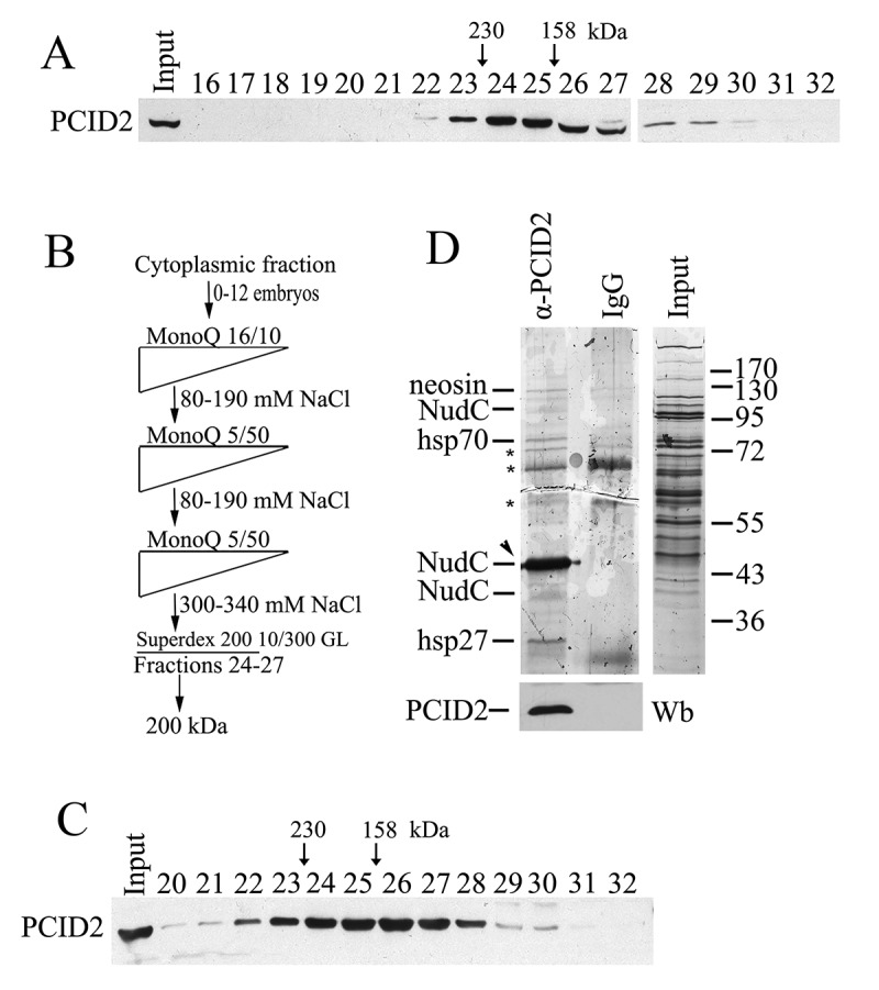
Figure 3.

PCID2 interacts with NudC in the cytoplasm
(a) The cytoplasmic extract from Drosophila embryos (0–12 h) treated with DNase I and RNase, was fractionated on a Superdex 200 10/300 GL gel filtration column, and the fractions were analysed for the presence of PCID2 by Western blot analysis. Fraction numbers are indicated. The void volume corresponds to fraction 10. (b) The schematic of purification of PCID2-interacting proteins from the cytoplasmic extract. At each step, the proteins were eluted with a NaCl gradient, and the fractions were analysed for the presence of PCID2 by Western blotting. The peak PCID2-containing fractions were collected and loaded into the next column. After fractionation on a Superdex 200 10/300 GL column, the material was loaded onto an immunosorbent with anti-PCID2 antibodies, washed, and eluted with acid glycine. (c) The migration profile of PCID2 on the Superdex 200 10/300 GL column at the final step of purification. (d) Preparation of PCID2-associated proteins purified from the cytoplasmic extract of embryos (silver staining). Fractions 23–28 (Fig. 3c) from the last step of fractionation were incubated with affinity-purified anti-PCID2 antibodies covalently bound to protein A Sepharose. Proteins were eluted, resolved by 9% SDS-PAGE, and analysed by mass spectrometry. The control immunoprecipitation of the same material with IgG and the protein extract (Input) are shown. The non-specific bands which were also found in IgG are indicated by asterisks. The preparation also contains the trace amounts of neosin, Hsp70, and Hsp26 which may specifically co-precipitate with PCID2. The amount of PCID2 in preparation was not high as it mostly remained bound to antibodies. The PCID2 band coincides with the NudC band (indicated by arrowhead) as both proteins have the same electrophoretic mobility in the gel. To confirm the presence of PCID2 the same preparations were stained with anti-PCID2 antibodies (bottom panel). The preparation also contains the additional weak bands of NudC which are likely NudC dimer (upper band) and the result of partial degradation or represent a different NudC isoform (lower band).
