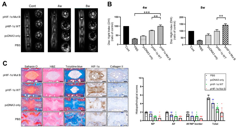
Figure 4.
CA HIF-1α (pHIF-1α Mut B) ameliorates disc degeneration. (A) Representative T2 weighted micromagnetic resonance imaging (MRI) of rat tail in the four groups at 4 and 8 weeks. (B) Disc height index (DHI) of four groups at 4 and 8 weeks. ** p-value < 0.01, *** p-value < 0.005. (C) Histology and immunohistochemistry in puncture-induced rat IVD degeneration model. Histological analysis of intervertebral discs by Safranin O, H&E, Toluidine blue staining, and immunohistochemical staining of HIF-1α and collagen II in the nucleus pulposus (NP) in all groups. Scale bars represent 100 μm. ** p-value < 0.01, *** p-value < 0.005.

