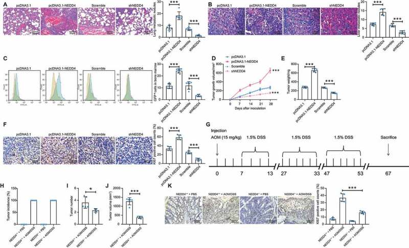
Figure 2.

Overexpression of NEDD4 promotes CC cell growth and metastasis in NOD/SCID mice. A-B, number of metastatic nodules in lung (A) and liver (B) tissues determined by HE staining; C, number of GFP-positive cells in mouse bone marrow determined by flow cytometry; D, growth curves of xenograft tumours in mice according to the tumour growth in vivo; E, weight of the xenograft tumours in mice; F, number of Ki-67 positive cells in tumour tissues determined by immunohistochemical staining; G, a diagram for CC induction by AOM/DSS in both WT C56J/6B mice (NEDD4+/+) and mut (NEDD4−/-) mice; H, occurrence rate of CC in mice following AOM/ASS of H2O treatment; I, number of tumours in each type of mice; J, total volume of the tumours in NEDD4+/+ and NEDD4−/- mice; K, rate of Ki67-positive cells in tumour tissues in both mouse types determined by immunohistochemical staining. There were 5 ~ 6 mice in each group. Data were exhibited as mean ± SD from three independent experiments. In panels A, B, C, E, F, H and K data were compared using one-way ANOVA, while data in panel D were determined by one-way ANOVA followed by Tukey’s multiple comparison test; and data in panels I and J were compared using unpaired t test. ** p < 0.01, *** p < 0.001
