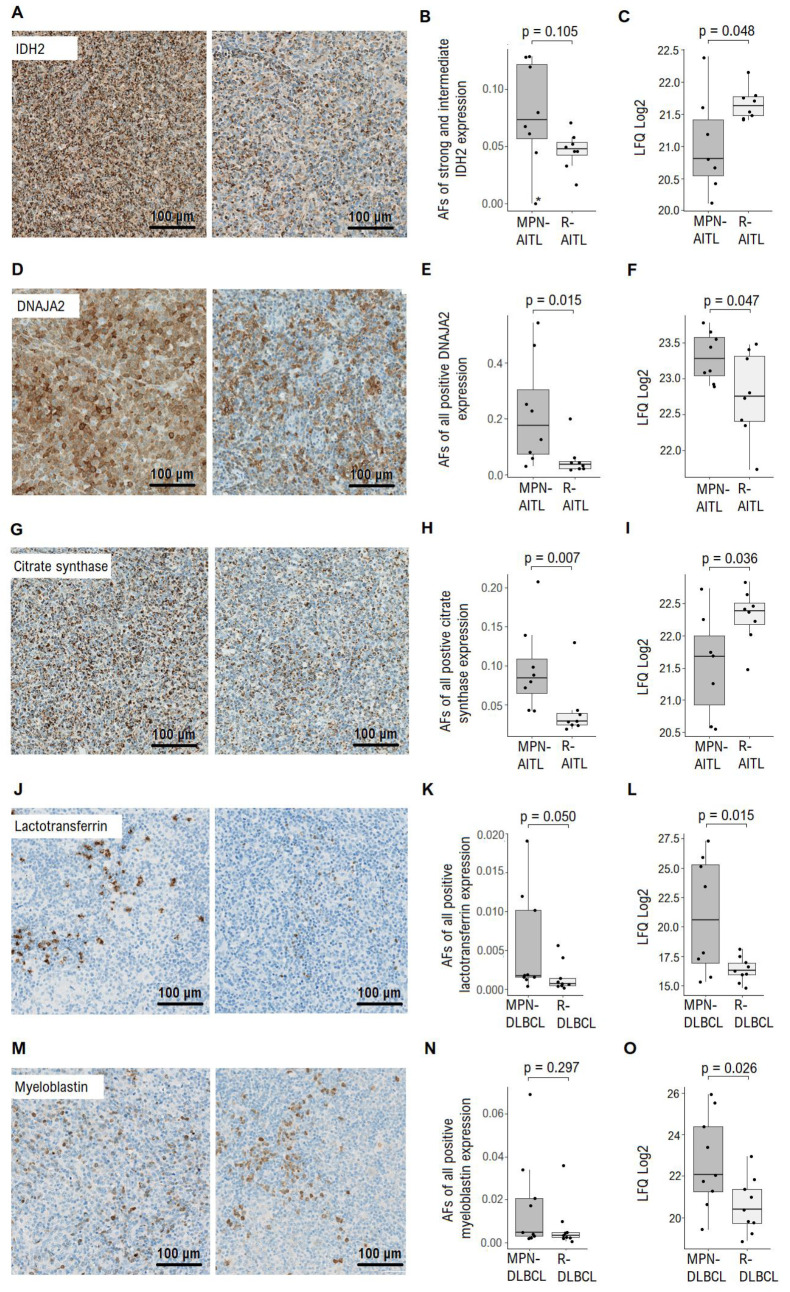
Figure 2.
Immunohistochemical evaluation of selected proteins. (A) Representative images of IDH2 protein staining expression (B) AFs of strong and intermediate intensity staining of IDH2 protein. * denote MPN-AITL patient with two known IDH2 gene mutations. (C) IDH2 protein expression identified by MS-based proteomics analysis. (D) Representative images of DNAJA2 staining expression. (E) AFs of all positive DNAJA2 protein staining. (F) DNAJA2 protein expression identified by MS-based proteomics analysis. (G) Representative images of citrate synthase staining expression. (H) AFs of all positive citrate synthase staining. (I) Citrate synthase protein expression identified by MS-based proteomics analysis. (J) Representative images of lactotransferrin staining expression. (K) AFs of all positive lactotransferrin staining. (L) Lactotransferrin protein expression identified by MS-based proteomics analysis. (M) Representative images of myeloblastin staining expression. (N) AFs of all positive myeloblastin staining. (O) Myeloblastin protein expression identified by MS-based proteomics analysis. Abbreviations: AF, area fraction; AITL, angioimmunoblastic T-cell lymphoma; DLBCL, diffuse large B-cell lymphoma; DNAJA2, DnaJ homolog subfamily A member 2; IDH2, isocitrate dehydrogenase 2; LFQ, label free quantification; MPN, myeloproliferative neoplasia; R-, reference sample.

