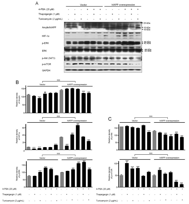
Figure 2.
The expressions of amylin/hIAPP, HIF1α, p-ERK, p-Akt, and p-mTOR proteins in pCMV-Entry-expressing cells and INS1E-hIAPP-overexpressing INS-1E cells under ER stress conditions. INS-1E cells were incubated in RPMI 1640 medium supplemented with 2% fetal bovine serum with/without thapsigargin (1 μM) for 6 h or tunicamycin (2 μg/mL) for 16 h and/or with/without 4-PBA (20 μM) at 37 °C with 5% CO2. Amylin/hIAPP, HIF1α, p-ERK, p-Akt, and p-mTOR were then analyzed by Western blot (A). The relative amounts of Amylin/hIAPP, HIF1α, and p-ERK (B) and p-Akt, and p-mTOR (C) were quantified as described in Materials and Methods. Data represent the mean ± SEM of three experiments. ** p < 0.01, *** p < 0.001 vs. 2% FBS in pCMV-Entry-expressing cells; ## p < 0.01, ### p < 0.001 vs. 2% FBS in INS1E-hIAPP-overexpressing cells; &&& p < 0.001, pCMV-Entry-expressing cells vs. INS1E-hIAPP-overexpressing cells.

