Figure 1.
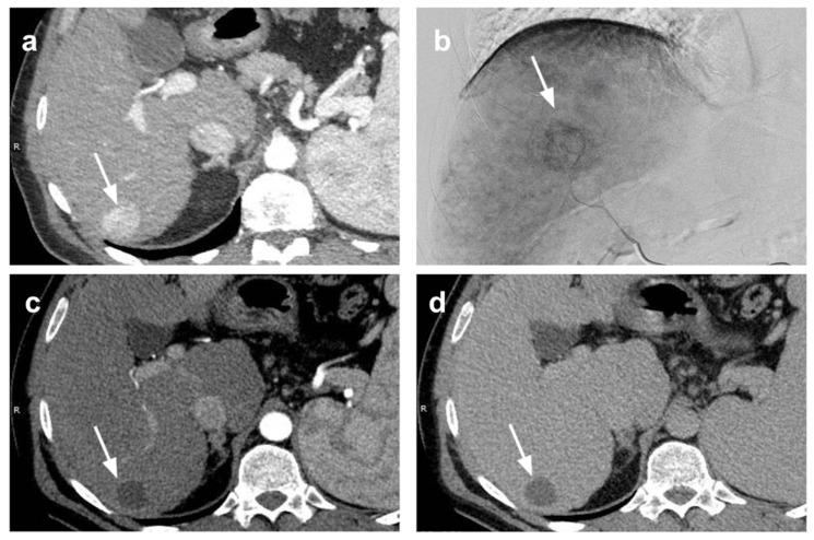
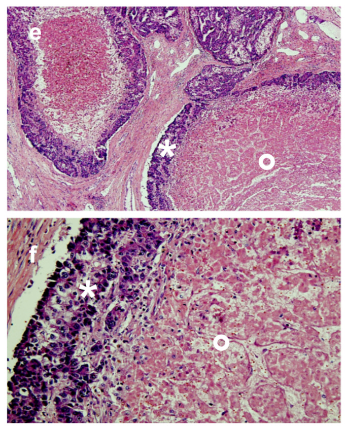
Bridging of HCC with TACE. Small HCC in hepatic segment 6 (arrow) at (a) pre-treatment CT scan in arterial phase and (b) at digital subtraction angiography. Complete radiological response was depicted at one-month CT evaluation after p-TACE (arrow) without appreciable enhancing tissue in (c) arterial and (d) portal-venous phase. The patient underwent liver transplantation two years after p-TACE. At pathologic examination of the explanted liver (e,f), extensive necrosis (*) was found in the treated area with presence of peripheral viable tumor tissue (ο) (e: magnification 20×; f: magnification 40×).


