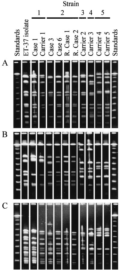
FIG. 1.
PFGE fingerprint analysis of the isolates. Isolates are identified by the same labelling as in Table 1 (except for “R. Case,” which stands for “remote case”). Standards were phage lambda concatamers. (A) Fingerprints generated with restriction endonuclease NheI; (B) fingerprints generated with restriction endonuclease SfiI; (C) fingerprints generated with restriction endonuclease SpeI. The results shown in each panel are from the same gel, and the lanes appear in the same order as on the original gel with the exception of those for remote case 2 (R. Case 2), which were moved from a position adjacent to the right-hand standard track for ease of comparison. To account for higher sample loading, the photographic contrast of the first two sample lanes of panel C (ET-37 isolate and Case 1) was adjusted independently of that for the rest of the gel in the preparation of this figure.

