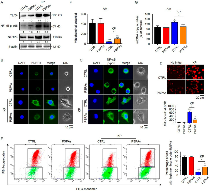
Figure 4.
PSPAs suppresses NLRP3 inflammasome activation and mitochondrial dysfunction induced by KP. (A) Immunoblotting of TLR4 and NLRP3 and phosphorylation of NF-κB p-p65 in PSPAs-treated and controlled AMs after treating with or without KP at an MOI of 20:1 for 6 h. Representative images of (B) NLRP3 (green) and (C) NF-κB p-p65 (green) and DAPI (blue).NF-κB p-p65 by laser confocal microscopy. (D) MitoSOX Red Mitochondrial Superoxide Indicator detected by fluorescence assay and mitochondrial potential measured by (E) JC-1 flow cytometry and (F) fluorescence assay. (G) Mitochondrial DNA copies were analyzed by qRT-PCR. Data (mean ± SEM) are representative of three independent experiments. One-way ANOVA (Tukey’s post-hoc); * p < 0.05, *** p < 0.001.

