Figure 3.
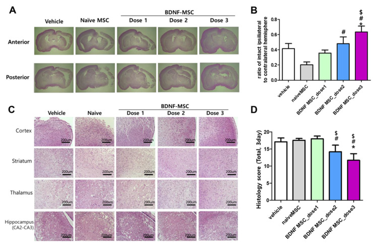
Morphological changes in the brains following hypoxic-ischemic brain injury and MSC transplantation. (A) Hematoxylin and eosin (H&E)-stained brain sections showing representative gross morphology of brains at 3 days after modeling. (B) The ratio of intact ipsilateral hemisphere to contralateral hemisphere was evaluated in the histologic brain section at 3 days after modeling in each group. (C) H&E-stained brain sections showing representative brain histology in cortex, striatum, thalamus, and hippocampus at 3 days after modeling. (D) Total sum of histologic scores in each area of brain; cortex, striatum, thalamus, and hippocampus. Data are expressed as mean ± standard error of the mean. * p < 0.05 compared to the HI + vehicle control, # p < 0.05 compared to the HI + naïve MSCs, $ p < 0.05 compared to HI-+ BDNF-eMSCs at dose 1 (approximately 1 × 10⁴ cells). Abbreviations: vehicle, HI + vehicle control, naïve MSCs; HI + naïve MSCs, BDNF MSC_dose1; HI + BDNF-eMSCs at 1 × 10⁴ cells dose; BDNF MSC_dose2; HI + BDNF-MSCs at 5 × 10⁴ cells dose; BDNF MSC_dose3; HI + BDNF-MSCs at 1 × 10⁵ cells dose.

