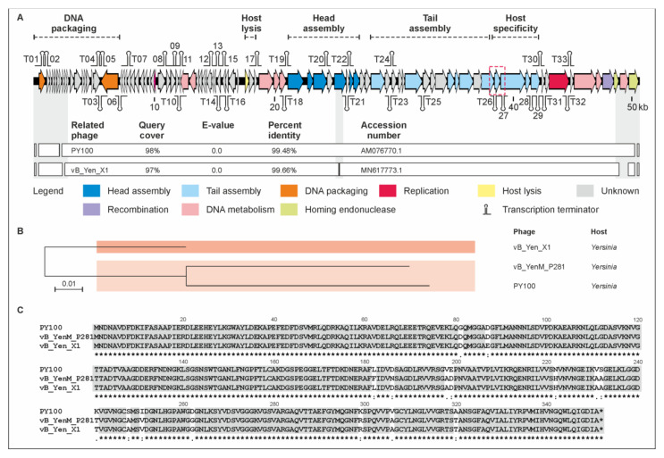
Figure 3.
Gene map of phage vB_YenM_P281 and its relationship with PY100 and vB_Yen_X1. (A) Gene map of vB_YenM_P281 and values of identity with the other two phages. White bars represent regions of high nucleotide similarity (>75%). (B) Phylogenetic tree of the phages. The scale bar represents the number of nucleotide substitutions per site. (C) Alignment of tail fiber proteins 1 (ORF 78 (PY100), 78 (P281) and 69 (vB_Yen_X1)). The location of the vB_YenM_P281 tail fiber gene is indicated in the gene map (red box).

