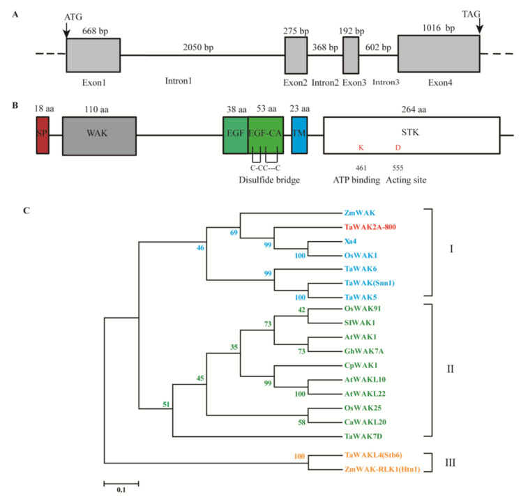
Figure 2.
Sequence and phylogenetic analyses of TaWAK2A-800. (A) Genomic structure of the TaWAK2A-800 gene. (B) Schematic diagram of the domain (shaded area) of the TaWAK2A-800 protein. The red box represents a signal peptide, the gray box represents the WAK-GUB domain, the green boxes represent EGF domains, the blue box represents the transmembrane domain, and the white box represents the STK domain. (C) Phylogenetic analysis of TaWAK2A-800 protein and 18 other WAK/WAKL proteins. The bootstrapped phylogenetic tree is constructed by using the neighbor-joining phylogeny of MEGA 7.0.

