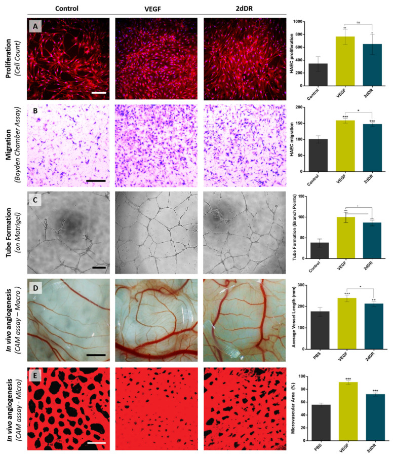
Figure 5.
Representative images demonstrating the pro-angiogenic potential of 2dDR compared to VEGF in terms of stimulating the (A) proliferation, (B) migration, (C) tube formation of HAECs, and in vivo angiogenesis assessed by (D) macro vessels and (E) microvasculature in ex ovo CAM bioassay. (*** p ≤ 0.001, ** p ≤ 0.01, * p ≤ 0.05, ns: not significant, n = 3). Scale bars represent 200 μm for proliferation, 250 μm for migration and tube formation images, 2 mm for macro and 50 μm for confocal microvessel images of CAM bioassay. Images, used in (A,D), are adapted from [32] with permission granted by Microvascular Research, Copyright 2020 Elsevier. Images, used in (E), is adapted from [30] with permission granted by Regenerative Medicine, Copyright 2019 Future Medicine.

