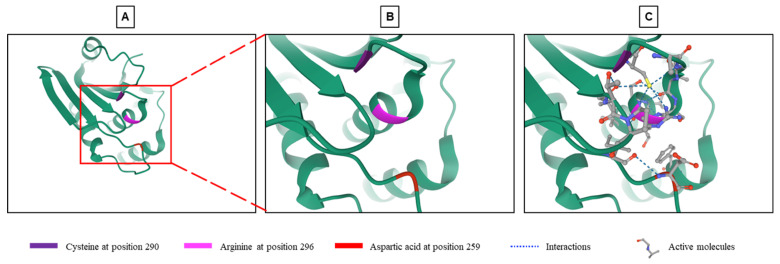
Figure 1.
Structure of the phosphatase active site of DUSP9. (A) Crystallographic representation of the phosphatase site of DUSP9 with a focus on its catalytic part (see region of interest in red box) composed of a cysteine at position 290, an arginine at position 296 and an aspartic acid at position 259 (from RCSB Protein Data Bank: https://www.rcsb.org/3d-view/3LJ8, accessed on 1 September 2021) [45]. (B) Higher magnification of region of interest shown in panel A. (C) Same image than in Panel B with inserted substrates and molecular interactions between the three amino acyls and substrate.

