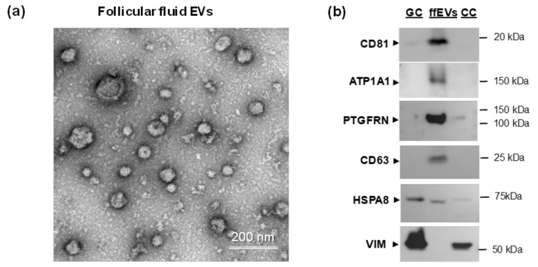
Figure 3.
Analysis of potential palmitoyl-proteins in follicular granulosa cells (GC), cumulus cells (CC) and small extracellular vesicles extracted from follicular fluid (ffEVs). (a) Representative transmission electron microscopy images of ffEVs. (b) Western blot analysis of CD81, ATP1A1, VIM and PTGFRN in bovine GC, CC and ffEVs. Tetraspanin CD63 and heat shock protein A8 (HSPA8) were used as known EV markers [54].

