Figure 2.
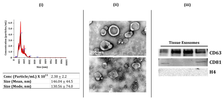
Characterization of human lung-tissue-derived Exosomes (i.e., exosome-enriched EVs. (i) Representative image for particle size distribution of lung-tissue-derived exosome in one sample as estimated using NanoSight NS300. Average particle size depicted as mean, mode, and particle concentration in lung-tissue-derived exosome samples (n = 3–5/group). (ii) Representative TEM images of lung-tissue-derived exosomes (n = 6). (iii) Immunoblot analysis of positive (CD63 and CD81) and negative (H4) exosomal markers derived from human lung tissue (n = 4).

