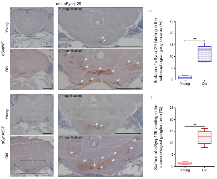
Figure 3.
Age-dependent increase of αSynp129 in αSyn-expressing flies. Representative immunohistochemistry paraffin-embedded brains stained with an anti-αSynp129 (EP1536Y) antibody on young (5 day-old) (a,c) or old (60 day-old) (b,d) flies expressing αSynWT (a,b) or αSynA53T (c,d). Magnification of area indicated in (a–d) are shown in (a’–d′). Note that localized staining of the αSynp129 is observed in the suboesophageal ganglion area as well as an increased detectability in older flies. White arrowheads indicate αSynp129 deposits. Scale bar represents 30 μm. Quantifications show the surface of αSynp129 labeling (EP1536Y antibody) in subesophageal ganglion area for αSynWT (e) or αSynA53T (f) in young (5- or 10-day-old) or old (50- or 60-day-old) flies. In the box and whisker plots in (e,f), boxes extend from the first to the third quartile, the line inside the boxes shows the median and the whiskers represent the min/max value of independent experiments: αSynWT (n = 4 for young, n = 6 for old) or αSynA53T (n = 5 for young, n = 6 for old). p-values of the group differences were calculated using Mann–Whitney test (** p = 0.0095 for αSynWT and ** p = 0.0043 for αSynA53T). Flies were raised on standard corn meal food supplemented with yeast.

