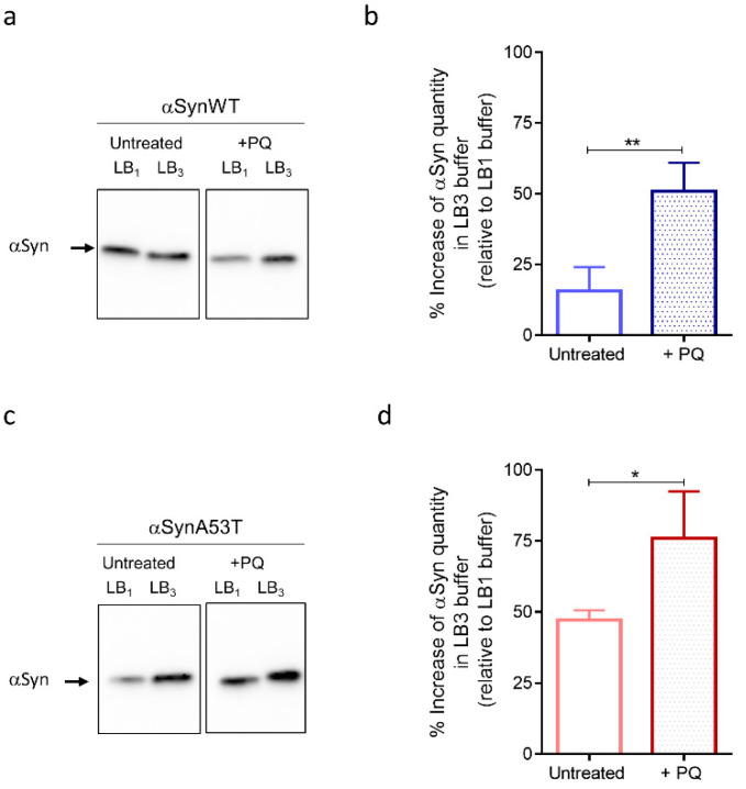
Figure 6.
Fly chronic exposure to PQ affects soluble αSyn detection in denaturating buffers. Western blot (antibody MJFR1) analysis on fly head extracts from control (untreated) or PQ exposed (+PQ) flies expressing αSynWT (a,b) or αSynA53T (c,d) realized at LT50. Heads (n = 100) were homogenized using a lysis buffer 1 (LB10.5% NP40) or a chaotropic lysis buffer 3 (LB3 urea/thiourea). The optical density of each sample was measured and normalized using a β-tubulin run on the same gel. The graphs are composites of six independent experiments (n = 3 for αSynWT and n = 3 for αSynA53T). The data, shown as mean with standard deviation in (b,d), represent the increase of the signal quantify with LB3 urea/thiourea compared to LB10.5% NP40 for a same experimental condition. p-values of the group differences were calculated using t-test (** p = 0.0045 for αSynWT and * p = 0.0203 for αSynA53T).

