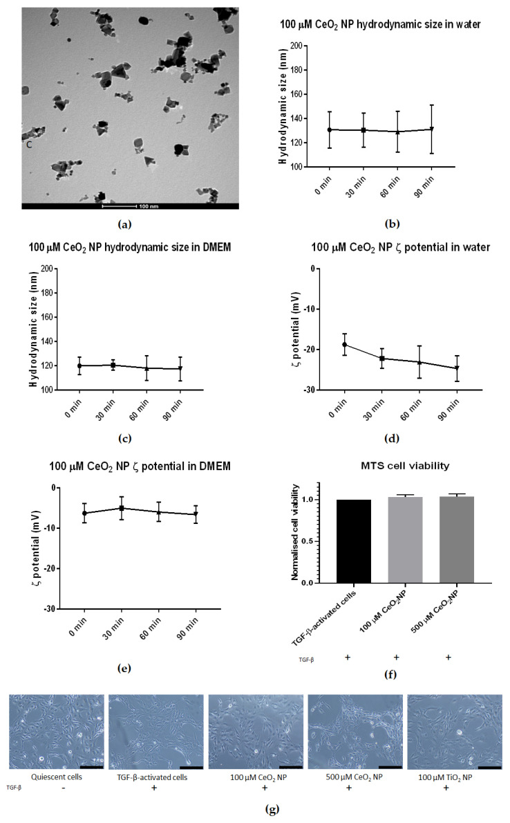
Figure 1.
Characterisation of CeO2 NP used in this study. (a) TEM analysis revealed CeO2 NP to be cubic-shaped and approximately 25 nm in diameter. (b) DLS analysis revealed CeO2 NP hydrodynamic size in water to be stable over 90 min at approximately 130 nm (n = 3). (c) CeO2 NP hydrodynamic size in 1% DMEM showed a slightly smaller size at approximately 120 nm and was also stable over 90 min (n = 3). (d) CeO2 NP in water showed a downward trend in ζ potential (n = 3). (e) CeO2 NP ζ potential in 1% DMEM showed more stable ζ potential (n = 3). (f) 100 µM and 500 µM CeO2 NP showed similar cell viability as TGF-β-treated control cells when measured using MTS assay (n = 3). (g) Light microscope images taken at 10× magnification of quiescent cells, TGF-β-activated cells, 100 µM CeO2 NP, 500 µM CeO2 NP and TiO2 NP-treated cells (n = 3). Scale bar represents 200 µm. Error bars represent standard error of the mean (SEM); one-way analysis of variance (ANOVA) was used to calculate significance.

