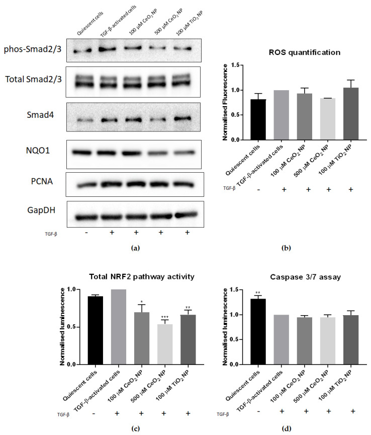
Figure 4.
CeO2 NP treatment reduces hallmarks of HSC activation. (a) Phosphorylated Smad2/3, total Smad2/3, Smad4 and NQO1 immunoblots. Phosphorylated Smad2/3 and Smad4 protein expression was decreased in 500 µM CeO2 NP-treated cells. Similarly, NQO1 expression was reduced in CeO2 NP-treated cells. PCNA expression showed no difference between TGF-β controls and CeO2 NP-treated samples. (n = 4) (b) ROS levels in CeO2 NP-treated cells were measured using flow cytometry. It can be seen that 500 µM CeO2 NP reduced oxidative stress to levels comparable to quiescent cell controls. (n = 3) (c) Nrf2 pathway activity. Total Nrf2 activity was measured using an ARE-promoter assay. The 100 µM CeO2 NP, 500 µM CeO2 NP and TiO2 NP-treated cells all showed significantly reduced Nrf2 activity. (n = 3) (d) Caspase 3/7 activity assay. CeO2 NP-treated cells showed no significant difference in caspase 3/7 activity compared to TGF-β controls. However, quiescent cells showed significantly higher caspase 3/7 activity. (n = 3). Error bars represent SEM; ANOVA was used to calculate significance. * p < 0.05, ** p < 0.01, *** p < 0.001.

